Theia is making tiny proteins easier to see. The laser is only part of the story.
Berkeley’s laser phase plate boosts electron-image contrast, with encouraging results for small proteins and bigger ambitions still ahead.
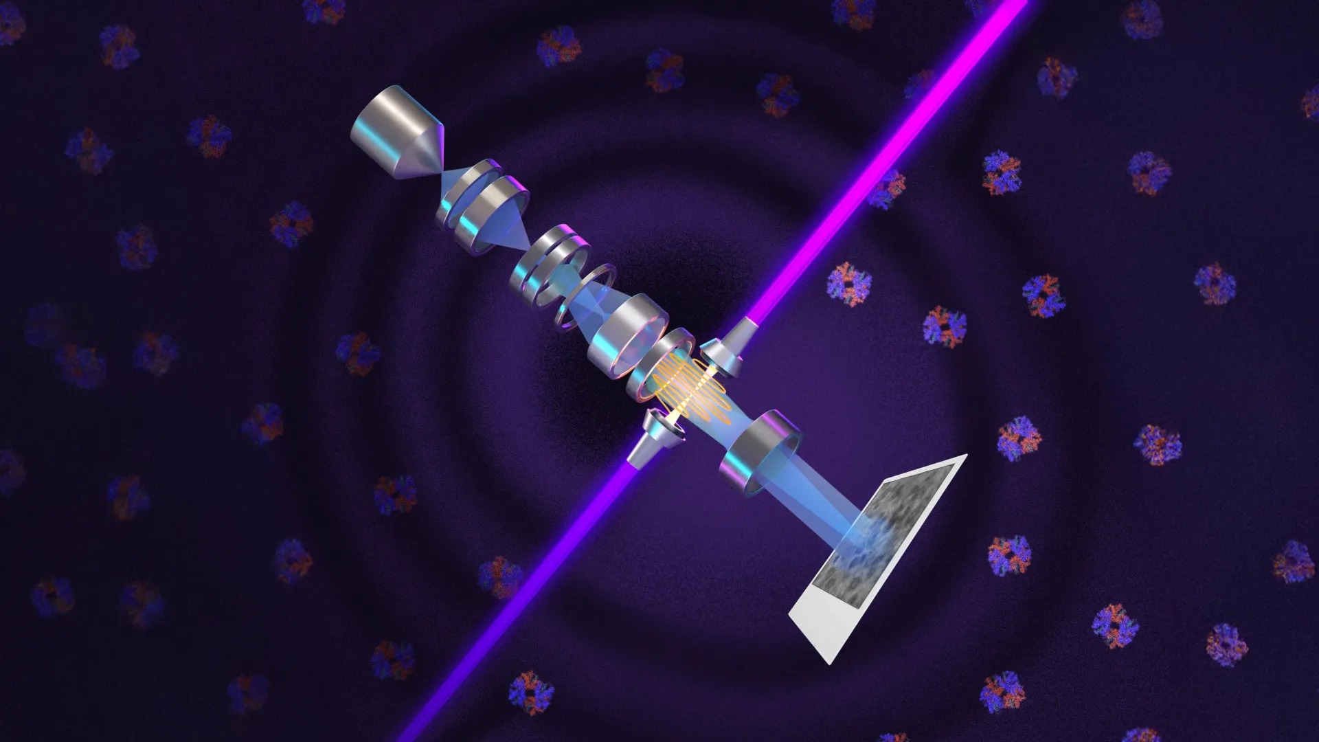
A powerful laser is helping scientists see proteins that are difficult to pick out with conventional cryoelectron microscopy. The innovation is not simply a bigger magnifying glass. It changes the contrast in an electron image, making faint structures easier to distinguish from their surroundings.
Research described by the University of California, Berkeley, and covered by ScienceDaily on October 22 uses a laser phase plate in a customized microscope called Theia. The team's reported demonstrations include improved images of hemoglobin, the oxygen-carrying protein in blood. Wider access to smaller proteins is the promise; a finished cure for a disease is not the result being announced.
Magnification is only half the problem
An image can be highly magnified and still be difficult to interpret. In biological microscopy, a specimen may produce only a weak difference between the structure a scientist wants to see and everything around it. Increasing contrast makes that difference easier to detect.
Phase-contrast microscopy addressed a version of this problem with light almost a century ago. Light changes phase as it passes through material, even when the change in brightness is difficult to notice. Converting phase differences into visible contrast made cellular structures easier to examine. Berkeley's work applies the underlying idea to electrons, using an intense laser rather than a conventional material phase plate.
What the laser actually changes
The laser interacts with the microscope's electron beam and shifts its phase. Highly reflective mirrors build up the light inside an optical cavity, concentrating enough intensity into a tiny region to produce the desired effect. The report describes approximately 75 kilowatts focused over a few micrometres.
That figure concerns the instrument's concentrated optical field. It should not be read as the power of a handheld medical device, or as a laser treatment administered to a patient. The technology belongs to a research microscope and a specialized imaging workflow.
Why hemoglobin is a useful test
Cryoelectron microscopy, usually shortened to cryo-EM, examines frozen specimens and combines information from many images to reconstruct molecular structures. It avoids the need to grow the crystals required for conventional X-ray crystallography, but smaller molecules remain challenging.
The Berkeley team tested several samples, including aldolase and hemoglobin. Aldolase is comparatively accessible to current cryo-EM methods; the smaller hemoglobin is closer to a difficult size range. Improvement in the harder case is important because it suggests the laser can help where ordinary images offer less usable signal—not merely make already easy samples look impressive.
A demonstrated threshold and a future ambition
The source describes imaging proteins as small as approximately 50 kilodaltons, although doing so remains challenging. A kilodalton is a unit of molecular mass, not a measure of image width. The report contrasts that with the difficulty conventional cryo-EM has below roughly 70 kilodaltons.
A further goal is to reach proteins around 17 kilodaltons, the size of myoglobin. That smaller target is an ambition, not an achieved limit in the account. Keeping those two numbers separate prevents a prospective improvement from becoming a false claim about what laboratories can routinely image now.
From isolated proteins to crowded cellular structures
The researchers also see an opportunity in cryoelectron tomography, or cryo-ET. This technique combines views from multiple angles to reconstruct three-dimensional arrangements. Its attraction is context: not just what an isolated molecule looks like, but where molecular machinery sits among other cellular structures.
At Biohub, a related design uses two lasers arranged at right angles, each operating at about half the power. The Berkeley findings appeared in Science; the related Biohub design was described in a preprint. Those publication statuses should not be treated as identical stages of independent review.
Better structural information can help scientists ask better questions about disease and drug targets. It does not eliminate sample preparation, radiation damage, computing requirements or the need to test a treatment in people. The immediate achievement is more fundamental: making a class of hard-to-see objects more accessible to measurement.
Why freezing is still part of the experiment
The cryo in cryo-EM refers to cold preparation conditions. Biological specimens are frozen for imaging; this is not a film of a living person's cells being examined inside their body. Descriptions of seeing molecular machinery in its cellular context should not be confused with keeping the imaged sample alive throughout the electron-microscopy measurement.
Electrons can damage a specimen, limiting how much information an experiment can collect. The source describes cooling and lower-dose imaging as key developments in the history of the technique. Better contrast helps extract information from a difficult image; it does not make the physical problem of radiation damage disappear.
This also explains why sample quality still matters. Freshness, preparation defects and bubbles can affect results. The researchers reported that the laser was particularly helpful for smaller particles and challenging specimens. That is a more precise claim than saying every image from every microscope will improve by one fixed percentage.
From a specialist instrument to a useful research tool
Theia is a customized instrument, and the report describes collaboration with microscope manufacturer Thermo Fisher Scientific. Building a strong result in one laboratory is different from making a technique straightforward to install, maintain and reproduce across many facilities.
The two-laser Biohub approach addresses practical issues including heat-related component failure and optical distortions. Engineering those details matters because a research method has to remain stable long enough to collect useful data, not merely work at one exceptional moment.
Berkeley's account places the effort on a timeline beginning with a laser-phase-plate proposal in 2010. The resulting machine combines a new component with decades of progress in electron optics, freezing methods, detectors and computation. Its appeal is not a shortcut past those technologies. It is a way of getting more useful contrast from the system they have made possible.